

Meta Title: Immunohistochemistry in Tumor Microenvironment Research
Meta Description: Learn how immunohistochemistry helps researchers map immune cells, protein biomarkers, and spatial relationships within the tumor microenvironment.
Suggested URL Slug: immunohistochemistry-tumor-microenvironment
Cancer research increasingly looks beyond tumor cells themselves. A tumor develops within a complex biological environment containing immune cells, fibroblasts, blood vessels, extracellular matrix, and signaling molecules. These components can influence tumor growth, metastasis, immune escape, and response to treatment.
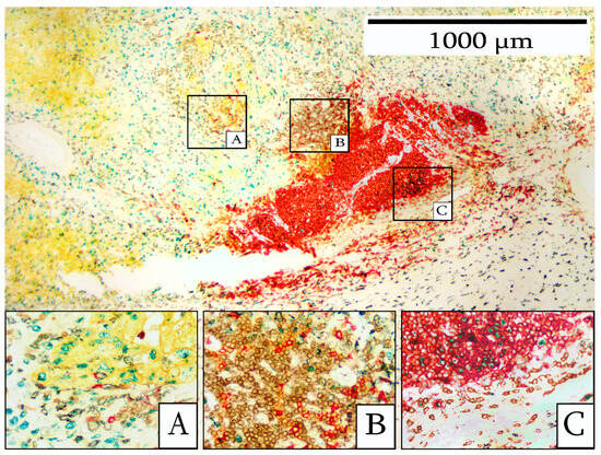
Understanding this environment requires methods that preserve spatial information. Immunohistochemistry (IHC) allows researchers to visualize specific proteins while retaining the architecture of the tissue. For studies requiring specialized staining and tissue analysis, immunohistochemistry services can support the investigation of protein expression and cellular distribution across tumor samples.
What Is the Tumor Microenvironment?
The tumor microenvironment, commonly abbreviated as TME, includes the malignant cells of a tumor and the surrounding non-malignant components.
Depending on the cancer, these may include:
- T lymphocytes
- B lymphocytes
- Macrophages
- Fibroblasts
- Endothelial cells
- Other stromal cells
- Extracellular matrix
These components interact through direct cellular contact and molecular signaling.
As a result, two tumors containing similar cancer cells may behave differently because their surrounding microenvironments differ.
Why Spatial Information Matters
Many molecular techniques require tissue to be disrupted before analysis.
RNA sequencing, for example, can provide extensive information about gene expression. Proteomic methods can characterize large numbers of proteins.
However, bulk measurements may not reveal exactly which cells produced a particular signal.
Suppose a tumor sample contains high levels of an immune-associated protein. Without spatial information, researchers may not know whether the protein originates from tumor cells, infiltrating immune cells, or surrounding stromal tissue.
IHC helps address this question by preserving tissue architecture while detecting selected molecular targets.
Identifying Immune Cells Inside Tumors
The immune composition of a tumor is an important area of cancer research.
Different immune populations may support or suppress antitumor responses depending on their phenotype, activation state, and location.
Researchers can use appropriate markers to investigate immune populations within tissue sections.
Instead of simply asking whether immune cells are present, tissue analysis can help answer more detailed questions:
- Are immune cells inside the tumor?
- Are they concentrated around the tumor boundary?
- Which immune populations are present?
- Do treated tumors show different infiltration patterns?
- Are immune cells located near particular tumor populations?
These spatial relationships can provide information that cell counts alone may miss.
Studying Tumor-Infiltrating Lymphocytes
Tumor-infiltrating lymphocytes, or TILs, are immune cells that have moved from circulation into tumor tissue.
T-cell populations receive particular attention because many cancer immunotherapies aim to stimulate or restore T-cell-mediated antitumor activity.
IHC can help researchers examine the distribution of lymphocyte-associated markers across tumor sections.
This makes it possible to distinguish between tumors with extensive immune infiltration and those where immune cells remain largely outside malignant regions.
Such patterns can help researchers characterize differences among tumor models and treatment groups.
Understanding Immune Checkpoint Biology
Immune checkpoints regulate immune responses and help prevent excessive immune activation.
Tumors can exploit some of these pathways to reduce antitumor immunity.
Research involving checkpoint proteins therefore often requires information about both expression and localization.
A protein may be present within a tumor sample, but understanding which cells express it can provide additional biological context.
Tissue-based analysis can help researchers investigate relationships among tumor cells, immune populations, and checkpoint-associated proteins within the same microenvironment.
Macrophages and the Tumor Environment
Macrophages represent another important component of many tumors.
These cells can exhibit diverse functional states and influence inflammation, tissue remodeling, angiogenesis, and immune responses.
Researchers may use tissue markers to examine macrophage abundance and distribution.
Spatial analysis can reveal whether macrophages are concentrated within tumor regions, near blood vessels, or in surrounding stromal tissue.
These patterns may provide clues about how macrophages interact with other components of the tumor microenvironment.
Cancer-Associated Fibroblasts
Immune cells are not the only important non-malignant cells in tumors.
Cancer-associated fibroblasts can influence extracellular matrix organization, signaling, tumor growth, and therapeutic response.
IHC can help researchers identify fibroblast-associated proteins and examine how these cells are distributed relative to malignant tissue.
This can be particularly valuable when investigating tumors with extensive stromal components.
Examining Tumor Vasculature
Tumors require access to nutrients and oxygen as they grow.
Formation and remodeling of blood vessels are therefore important areas of cancer biology.
Researchers can use endothelial and vascular markers to examine blood-vessel distribution within tumors.
Tissue analysis may help quantify vascular density or investigate whether experimental treatments alter tumor-associated vasculature.
Combining vascular markers with tumor and immune markers can provide a broader view of how different components of the microenvironment interact.
Why Biomarker Location Can Matter as Much as Abundance
Biomarker research often focuses on whether expression is high or low.
In heterogeneous tissue, location can be equally informative.
Consider a protein detected at similar overall abundance in two tumor samples. In the first, expression might occur mainly on tumor cells. In the second, it could be concentrated in immune cells.
A bulk measurement could make these samples appear similar.
IHC can reveal that they represent different biological patterns.
This illustrates why spatial context can add another dimension to biomarker research.
Multiplex Approaches Expand Tissue Analysis
Traditional IHC frequently examines individual markers on separate tissue sections.
Multiplex methods can detect several targets within the same sample.
This enables researchers to investigate multiple cell populations while preserving their spatial relationships.
A multiplex experiment might simultaneously examine markers associated with:
- Tumor cells
- T cells
- Macrophages
- Stromal cells
- Immune regulation
Researchers can then analyze not only how many cells of each type are present but also how close different populations are to one another.
Digital Pathology Makes IHC More Quantitative
Tissue analysis has traditionally relied heavily on visual microscopic assessment.
Digital pathology is expanding the quantitative possibilities.
Whole-slide scanners can convert stained slides into high-resolution digital images. Image-analysis software can then assist with measurements such as:
- Positive-cell percentage
- Staining intensity
- Cell density
- Tissue segmentation
- Spatial distribution
These approaches can be particularly useful when researchers need to compare large numbers of samples consistently.
Human review remains important, especially when tissue morphology is complex or automated segmentation performs poorly.
Antibody Validation Remains Essential
Sophisticated imaging cannot compensate for an antibody that does not reliably recognize its intended target.
Researchers should evaluate antibody specificity and suitability for the tissue, species, fixation method, and application.
Appropriate positive and negative controls are also important.
Unexpected staining patterns should be investigated rather than automatically interpreted as biological findings.
This is particularly important in multiplex experiments, where several antibodies and detection systems may interact within the same workflow.
Using IHC to Study Treatment Response
Tissue analysis can also help researchers investigate how the tumor microenvironment changes following experimental treatment.
For example, researchers may compare treated and untreated tumors to determine whether therapy changes:
- Tumor proliferation
- Cell death
- Immune infiltration
- Checkpoint expression
- Macrophage distribution
- Vascular characteristics
Using immunohistochemistry services for such studies can provide access to tissue processing, staining, imaging, and analysis capabilities when these workflows are not available internally.
These measurements can complement tumor-growth data and other molecular assays.
Combining IHC With Other Technologies
No single method fully describes the tumor microenvironment.
Researchers increasingly combine tissue imaging with:
- Flow cytometry
- RNA sequencing
- Single-cell sequencing
- Proteomics
- Genomic analysis
Each technology provides a different perspective.
Single-cell sequencing can characterize molecular states across thousands of individual cells, while IHC can reveal where selected populations are located within intact tissue.
Integrating these methods can produce a more complete view of tumor biology.
Looking Ahead
Cancer research is moving from treating tumors as uniform masses toward understanding them as complex biological ecosystems.
Immunohistochemistry remains valuable in this transition because it connects molecular markers with cells and tissue architecture.
Multiplex staining, digital pathology, spatial analysis, and computational imaging are increasing the amount of information researchers can obtain from a single tissue section.
As cancer therapies become more targeted and immune-focused, understanding where biomarkers and immune populations occur within tumors may become just as important as knowing whether they are present at all.
